Views
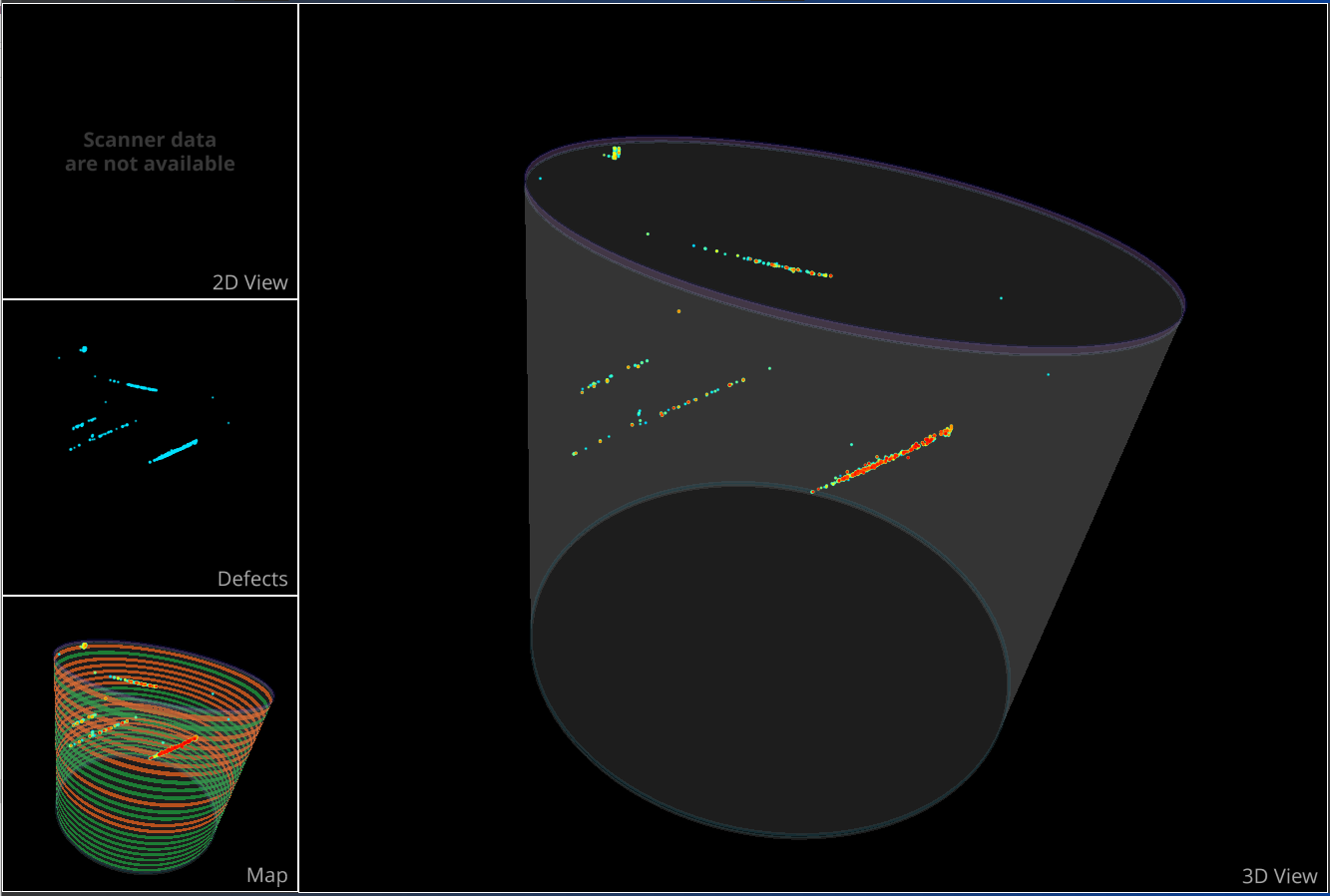
The views panels is divided into four subview as described below. Main view can be selected with a double click on desired sub view.
The three former view are 3D environment views with an Arcball camera that can be manipulated using:
LMB: to orbits the view around camera center point.
RMB or scroll: to zoom in/out focusing camera center point.
SHIFT + LMB: to translate the camera center point
CTRL + RMB: to rotate around camera center point
3D View
The 3D view contains the full 3D model with defects inside and auxiliary element for context (boundaries, exclusion zone, etc.).
Visibility and color of the auxiliary element can be changed with Crystal Module
Visibility and color of the defects element can be changed with Defects Module or using right click on it.
Defects
The defects view contains only the selected defects in Defects Module which are visible.
Map
Map view contains the full 3D model with in overlay the mapped items (i.e. wafers). Wafer appears by default in green if they are considered as “good material”, in red in case of “bad material” and in gray if the item is not fitting inside the crystal shape (if it overflows).
Visibility and color of the auxiliary element can be changed with Crystal Module
Visibility and color of the defects element can be changed with Defects Module or using right click on it.
Visibility, color and all processing setting about mapping can be set up in the Mapping Module.
2D View
2D view is here to navigate through raw tomography images if they are available. If no data are available the message “Scanner data are not available” is displayed
Note
SVCI images are raw data produced by Scientific Visual equipments and are usually not available without a machine. For optimization reasons the SVCI are not included directly into SVCM 3D model. You need at least to have access to a valid SCAN folder structure.
The control of this view are slightly different because the camera is now a panzoom camera which allow optimized 2D displays and can be manipulated using:
LMB: to translate the camera center point.
RMB or scroll: to zoom in/out focusing camera center point.
CTRL + scroll or dedicated button: to scroll through tomography images.